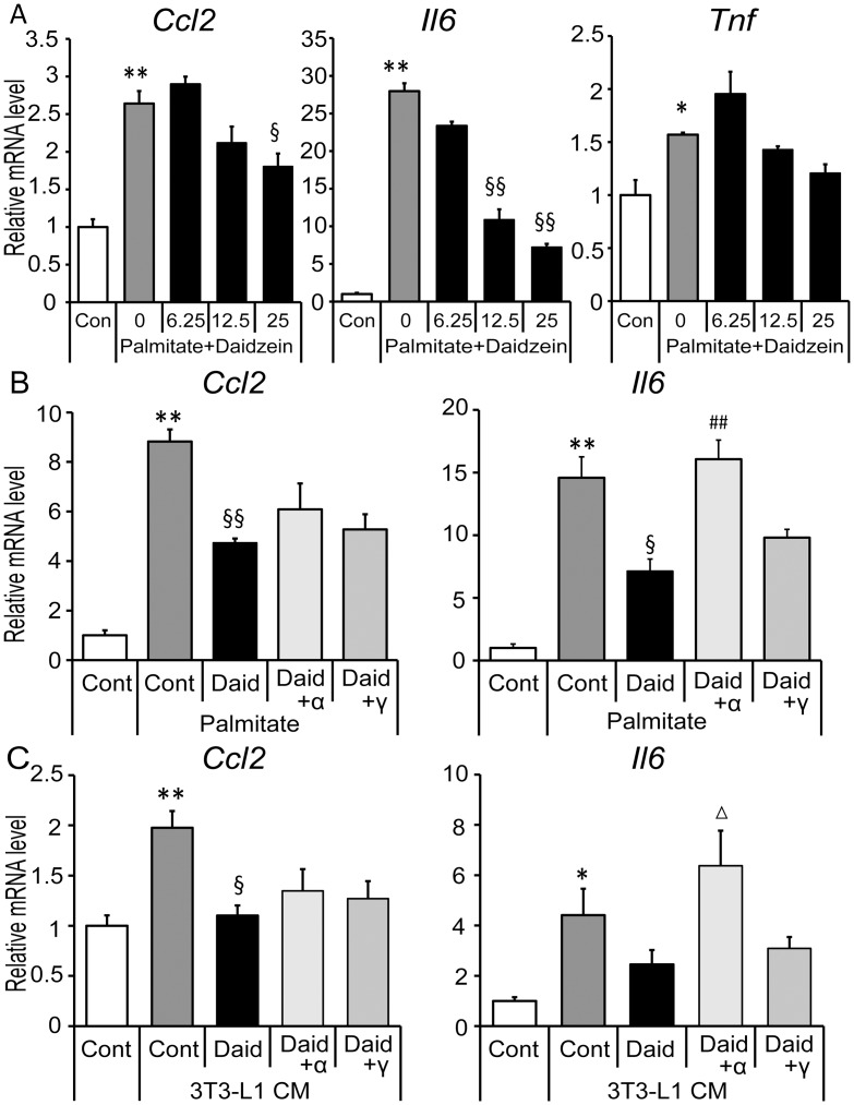
Fig 4. Effects of daidzein via PPAR-α/γ inhibition in palmitate- and CM- treated macrophages.
(A) RAW264 macrophages were cultured in the serum-free medium with the indicated concentrations (6.25–25 μM) of daidzein for 1 h, then stimulated with 400 μM palmitate for 4 h. (B) Cells were cultured in serum-free medium with DMSO (Cont), 25 μM of daidzein (Daid), Daid + 10 μM of GW6471 (Daid+α) or Daid + 10 μM of GW9662 (Daid+γ) for 1 h, then 400 μM palmitate was added and cells were incubated for another 4 h. (C) Cells were cultured in CM derived from hypertrophied adipocytes with Daid, Daid+α or Daid+γ for 24 h. Messenger RNA levels of each gene were quantified by real-time PCR and normalized to β-actin expression. Values are expressed as the fold change compared with the vehicle control that was arbitrarily set to 1. *, p<0.05; **, p<0.01 versus Cont. §, p<0.05; §§, p<0.01 versus palmitate Cont or CM Cont. Δ, p<0.1; ##, p<0.01 versus palmitate Daid or CM Daid.

