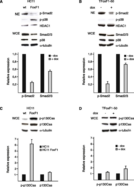
Fig. 5.

FoxF1 represses Smad2 activity. a, upper panel, western blot analysis of nuclear extracts (NE) from HC11 and HC11FoxF1 cells probed with phosphospecific Smad2 and p38 antibodies, stripped and reprobed with HDAC-1 antibody. Total levels of Smad2/3 and p38 were analyzed in whole cell extracts (WCE). b, upper panel, western blot analysis of nuclear extracts from TFoxF1-50 cells, untreated or dox-treated for 24 h, probed as in (a). c, upper panel, western blot analysis of whole cell extracts from HC11 and HC11FoxF1 cells probed with phosphospecific p130Cas antibody, stripped and re-probed with p130Cas and α-tubulin antibodies. d, upper panel, western blot analysis of whole cell extracts from TFoxF1-50 cells untreated or dox-treated for 24 h, probed as in (c). a-d, lower panels show densitometry
