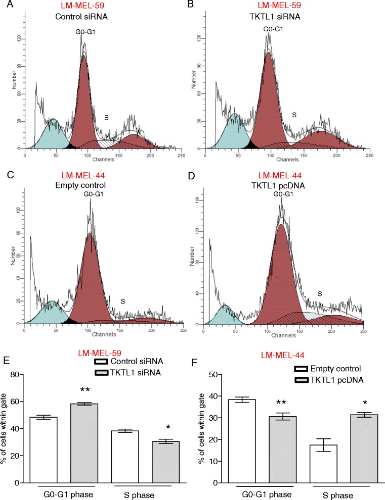
Fig. 4.

Loss of TKTL1 expression changes cell cycle distribution of melanoma cells. Cell cycle phases were determined by propidium iodide staining of melanoma cells and subsequent flow cytometric analysis. A representative histogram of cell cycle analysis of LM-MEL-59 is shown after a control siRNA treatment and b TKTL1 siRNA treatment. Analysis of percentage of cells in e G0-G1 phase and S phase cell after treatment of LM-MEL-59 with TKTL1 or control siRNA. Values are ± SD of three experiments in triplicate (*, p < 0.05, **, p < 0.005). Histograms depicting distribution of cell cycle phase in LM-MEL-44 after treatment with c empty vector control and d TKTL1 pcDNA. Cell cycle distribution of f GO-G1 and S phase cell population after 48 h of ectopic expression of TKTL1 or empty vector in LM-MEL-44 was performed. Values are ± SD of three experiments in triplicate (*, p < 0.05, **, p < 0.005)
