
Fig. 1.
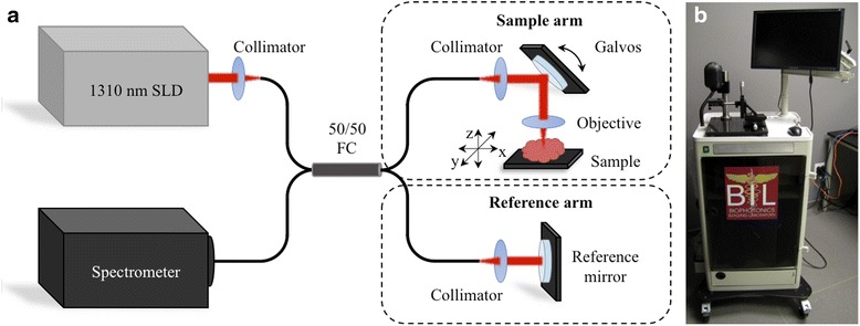
OCT system design for intraoperative assessment of ex vivo human lymph nodes for metastasis. a Schematic diagram and b photo of the OCT system. The red arrows indicate the path of near-infrared light travels along the optical fibers from the superluminescent diode (SLD) source, through the 50/50 fiber coupler (FC), splitting between the sample arm or reference arm. The reflected light from the tissue sample and the reference mirror is collected by the optical system, and travels back through the optical fibers and 50/50 FC to the line-scan detector in the spectrometer, the signal output of which was used to calculate the 3D-OCT images