
Fig. 2.
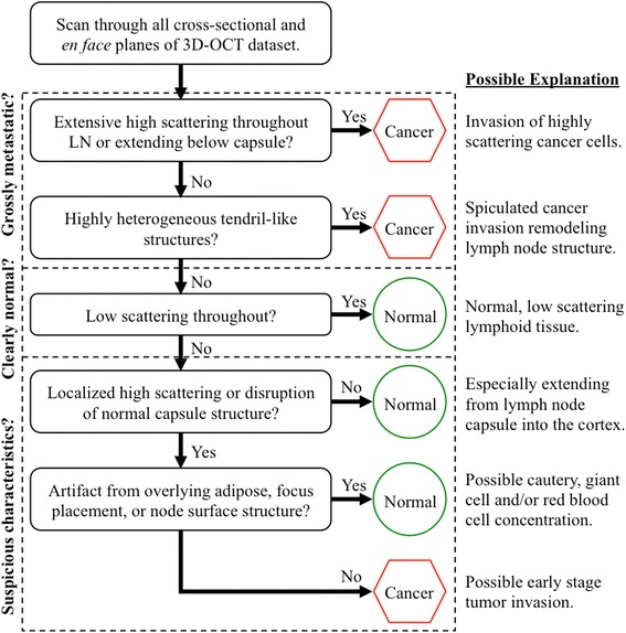
The decision tree diagram used for analyzing 3D-OCT images. A brief, sample training image set and this decision tree were developed for providing the blinded readers with direction for identifying native and abnormal lymph node anatomical structure, as well as possible image artifacts from imaging limitations in surgery. Each trained reader classified the OCT datasets as either “metastatic” or “non-metastatic”