
Fig. 4.
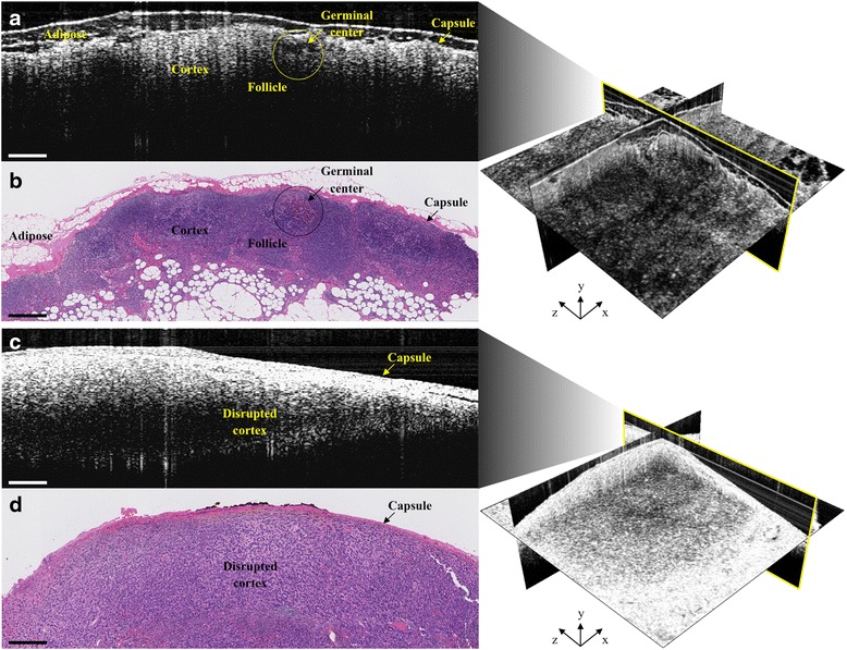
Representative intraoperative OCT (a & c) and corresponding histopathology (b & d) images of a normal, non-metastatic (top) and cancerous, metastatic (bottom) human lymph node. In a & b, normal lymph node structures, such as the capsule, cortex, follicles and germinal centers, as well as adipose, can be identified in both images. In c & d, metastatic invasion of cancer cells disrupts the normal lymph node cortex structure and can disrupt identification of follicles and germinal centers. All scale bars: 0.5 mm