
Fig. 5.
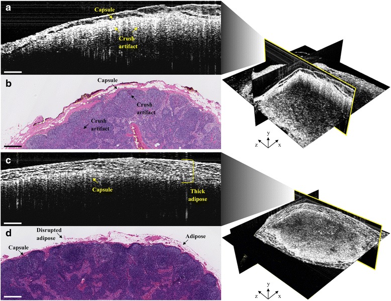
Representative intraoperative OCT (a & c) and corresponding histopathology (b & d) images of false positive (top) and false negative (bottom) cases. False positives can result from crush artifact, which mimics the bright white (high) OCT signal intensity of metastatic cancer cell invasion (Fig. 4c). False negatives can result from thick overlying adipose, which reduces imaging depth penetration, affects optical beam quality and resolution, and, consequently, underlying lymph node signal intensity. Some of the overlying adipose in the histology (d) is missing, most likely disrupted and/or lost during tissue processing. All scale bars: 0.5 mm