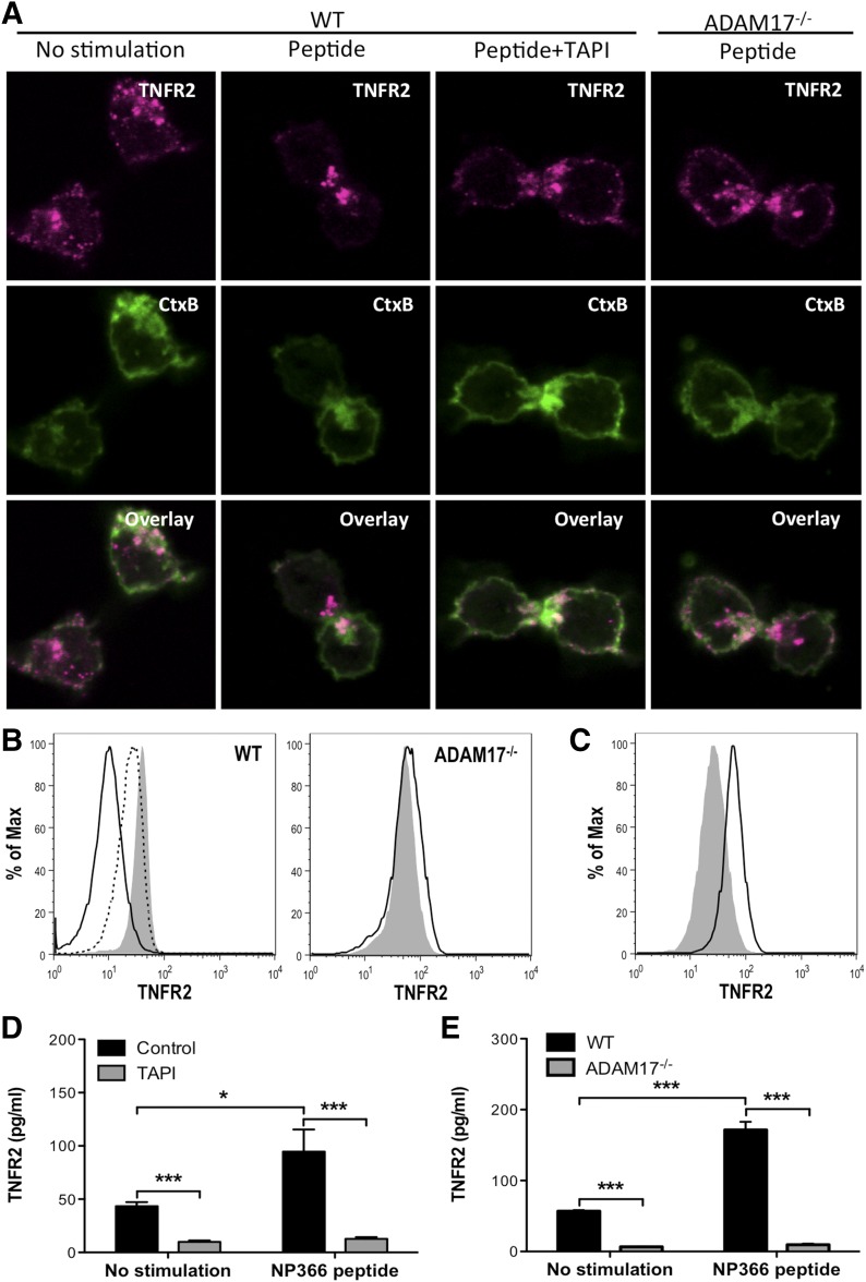
Figure 4. ADAM17 is required for TNFR2 shedding by influenza-specific CD8+ T cells.
TNFR2 shedding was examined in vitro on NP366–374-specific CD8+ T cells with NP366–374 peptide stimulation. (A) TNFR2 expression and CtxB binding were examined on unstimulated or peptide-stimulated and TAPI-treated, peptide-stimulated WT CD8+ T cells after 15 min of stimulation by confocal microscopy. Alternatively, TNFR2 and CtxB expression was examined on ADAM17−/− CD8+ T cells after 15 min of stimulation. (B) Representative histograms of memTNFR2 expression on unstimulated (gray), peptide-stimulated (black line), or TAPI-treated, peptide-stimulated (dashed line) WT or ADAM17−/− CD8+ T cells after 15 min of stimulation. (C) Unstimulated WT CD8+ T cells were left alone (gray) or treated with TAPI, and memTNFR2 expression was assessed 4 h later by flow cytometry. (D) Effects of TAPI on constitutive and peptide-stimulated solTNFR2 production by WT CD8+ T cells after 1 h of stimulation, as measured by ELISA. (E) Constitutive and peptide-stimulated solTNFR2 production by WT or ADAM17−/− CD8+ T cells after 1 h of stimulation, as measured by ELISA. Data represent means ± sd. Data are representative of 3 independent experiments with each condition conducted in triplicate. *P < 0.05; ***P < 0.005.

