
Figure 4. Ito kinetics are altered in chronic HF.
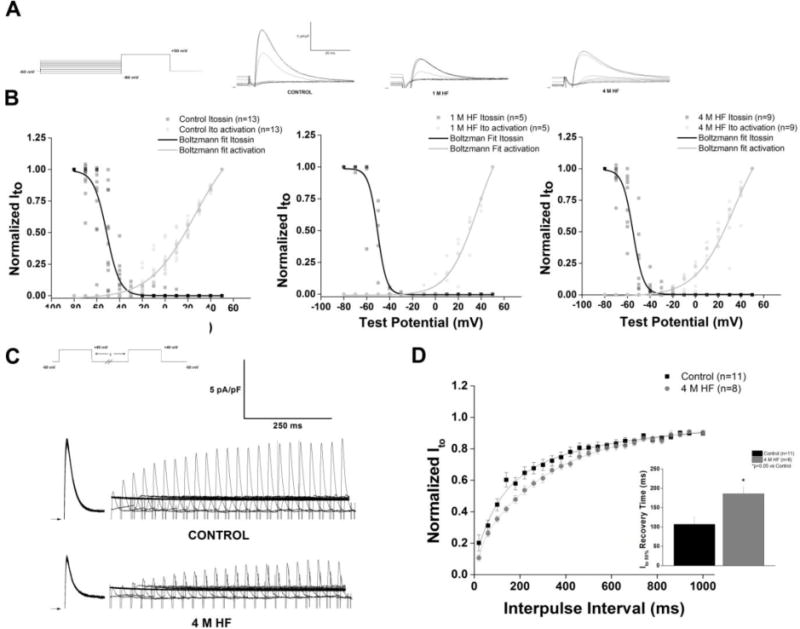
A. Representative steady state inactivation traces of Ito recorded with the voltage protocol displayed in the inset. B. Steady state inactivation and activation curves of Ito fit to Boltzmann functions, demonstrates a “window” current, that is reduced as the heart fails C. Representative traces elicited by two-step protocol in control and 4 M HF; voltage protocol in inset. D. Summary data of recovery from inactivation; HF significantly prolongs recovery to 50% of total Ito current (p<0.05 vs control).