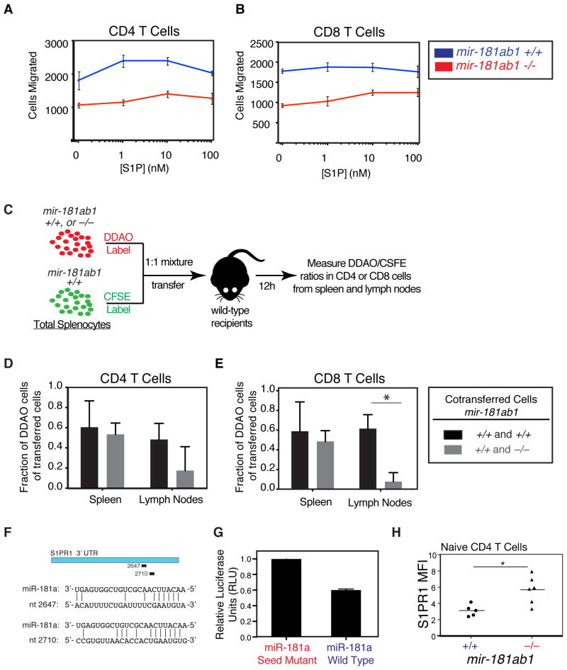
Figure 7. Effects of loss of mir-181a-1/b-1 on chemotaxis to sphingosine-1-phosphate.
(A–B) Splenocytes from mir-181a-1/b-1-knockout and wild-type mice were placed in transwells above varying concentrations of S1P and allowed to migrate for 3 h at 37 °C. Migrated CD4 and CD8 T cells were counted by flow cytometry. Number of (A) CD4 and (B) CD8 T cells migrated after 3 h in response to various concentrations of S1P (error bars are SD; *, p < 0.05, unpaired t-test). (C) Schematic depicting in vivo migration assay. CFSE-labeled splenocytes from wild-type mice were mixed at a 1:1 ratio with DDAO-labeled wild-type or mir-181a-1/b-1-knockout splenocytes and co-transferred into wild-type recipients. Spleens and lymph nodes were collected 12 hours after transfer and the percentages of DDAO-labeled cells in total transferred (D) CD4 and (E) CD8 T cells in spleens and lymph nodes were measured (bars are SD). (F) Putative miR-181a recognition sites in S1PR1 3′ UTR and base pairings with miR-181a as predicted by miRanda (positions are relative to start of 3′ UTR). (G) Relative luciferase levels in the presence of miR-181a or miR-181a seed-mutant. The S1PR1 3′ UTR was cloned into a luciferase reporter construct and co-transfected into BOSC cells with miR-181a wild-type and seed-mutant controls. Units were normalized to an internal Renilla luciferase control and to seed-mutant control (bars are SD).

