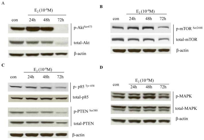
Figure 5. E2 degraded Akt-associated proteins.
(A) Regulation of Akt by E2. MCF-7:5C cells were treated with vehicle (0.1% EtOH) or E2 (10−9 mol/L) for different time points indicated. Cell lysates were harvested. p-Akt and total Akt were examined by Western blotting. β-actin was measured as loading control. (B) Regulation of mTOR by E2. Cells were treated the same as in (A). p-mTOR and total mTOR were examined by Western blotting. β-actin was measured as loading control. (C) Regulation of PTEN and p85 by E2. Cell lysates were the same as above. p-PTEN, total PTEN, p-p85, and total p85 were examined by Western blotting. β-actin was measured as loading control. (D) Regulation of MAPK by E2. MCF-7:5C cells were treated with vehicle (0.1% EtOH) or E2 (10−9 mol/L) for different time points indicated. Cell lysates were harvested. p-MAPK and total MAPK were examined by Western blotting. β-actin was measured as loading control.

