Figure 1.

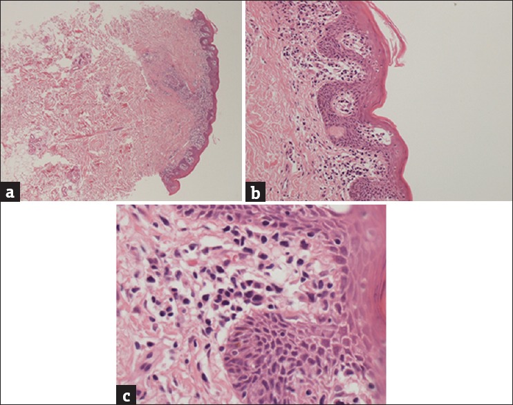
Biopsy demonstrating dermal and follicular infiltrate (H and E, ×10) (a) with epidermotropism and Pautrier microabscess (H and E, ×40) (b) and mycosis fungoides cells with cerebriform nuclei (H and E, ×100) (c)

Biopsy demonstrating dermal and follicular infiltrate (H and E, ×10) (a) with epidermotropism and Pautrier microabscess (H and E, ×40) (b) and mycosis fungoides cells with cerebriform nuclei (H and E, ×100) (c)