Figure 2.

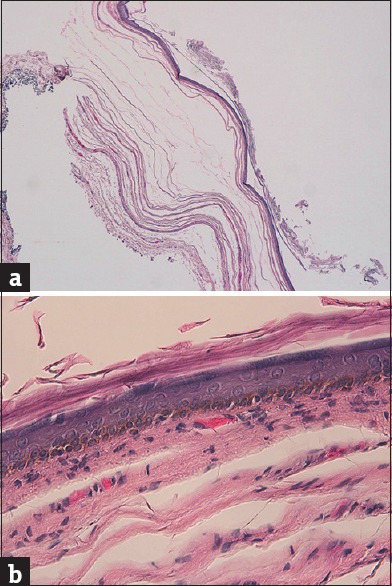
(a) Histological examination showed a cystic lesion containing abundant laminated keratin (H and E, ×2.5). (b) Walls were formed by keratinizing epithelium with the presence of granular layer (H and E, ×40)

(a) Histological examination showed a cystic lesion containing abundant laminated keratin (H and E, ×2.5). (b) Walls were formed by keratinizing epithelium with the presence of granular layer (H and E, ×40)