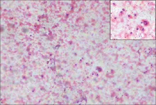
Figure 3.

Fine needle aspiration cytology (H and E) ×100 shows pleomorphic cells arranged in small clusters, groups, and individually scattered. Inset shows large pleomorphic centrally placed, hyperchromatic nucleus with abundant cytoplasm. Some of the cells showing eccentrically placed nucleus and prominent nucleoli. Few binucleated cells are also seen
