Figure 5.

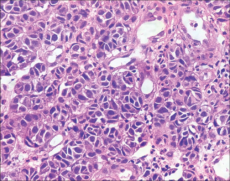
Histopathology of skin H and E stain ×400. Cuboidal cells with hyperchromatic nuclei and moderate amount of foamy cytoplasm and few cells showing eccentric nuclei and pleomorphism and mitotic activity

Histopathology of skin H and E stain ×400. Cuboidal cells with hyperchromatic nuclei and moderate amount of foamy cytoplasm and few cells showing eccentric nuclei and pleomorphism and mitotic activity