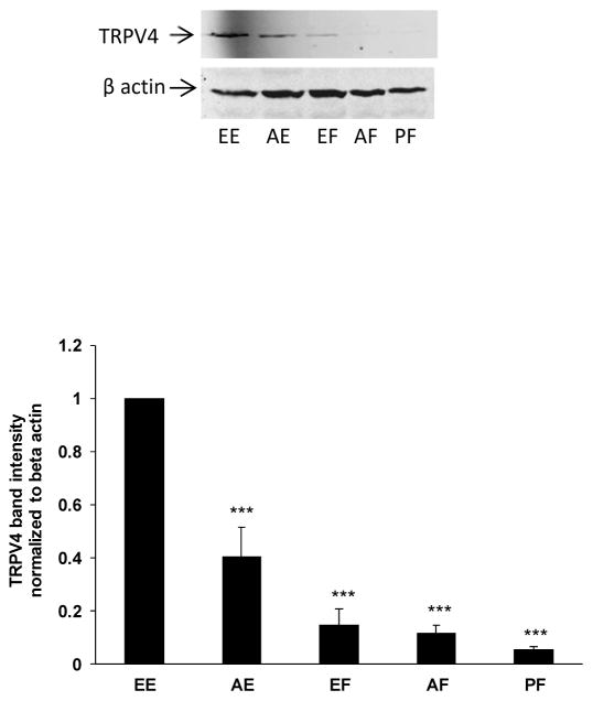
Fig. 5.
Western blot showing TRPV4 protein detected in different regions of the epithelium and cortical fiber mass. The different lens regions are: EE equatorial epithelium; AE anterior epithelium; EF equatorial fibers; AF anterior fibers; PF posterior fibers. The typical western blot shows TRPV4 (upper) and β-actin (lower), which was used as a loading control. The bar graph shows densitometric analysis of TRPV4 band density normalized to β-actin. The results are the mean ± SEM of data from 3 different lenses. ***(p<0.01) indicates a significant difference compared to band density observed in the equatorial epithelium.

