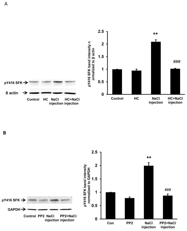
Fig. 7.
The influence of HC067047 (panel A) or PP2 (panel B) on SFK activation in the epithelium of lenses that received 5 μl of hyperosmotic NaCl solution injected ~1 mm beneath the posterior pole. Lenses were preincubated with or without 10 μM HC067047 (20 min), or 10 μM PP2 (40 min), subjected to the injection maneuver, then incubated for a further 5 min in the continued presence or absence of HC067047 or PP2, respectively. Control lenses did not receive an injection. On the left of each panel the typical western blot shows pY416-SFK (upper) and β-actin (lower) which was used as loading control. The bar graphs show densitometric analysis of pY416-SFK normalized to β-actin. The results are the mean ± SEM of data from 3 independent experiments. ** (p<0.01) indicates a significant difference compared to control and ### (p<0.001) indicates a significant difference compared to the NaCl injection group.

