Fig. 2.
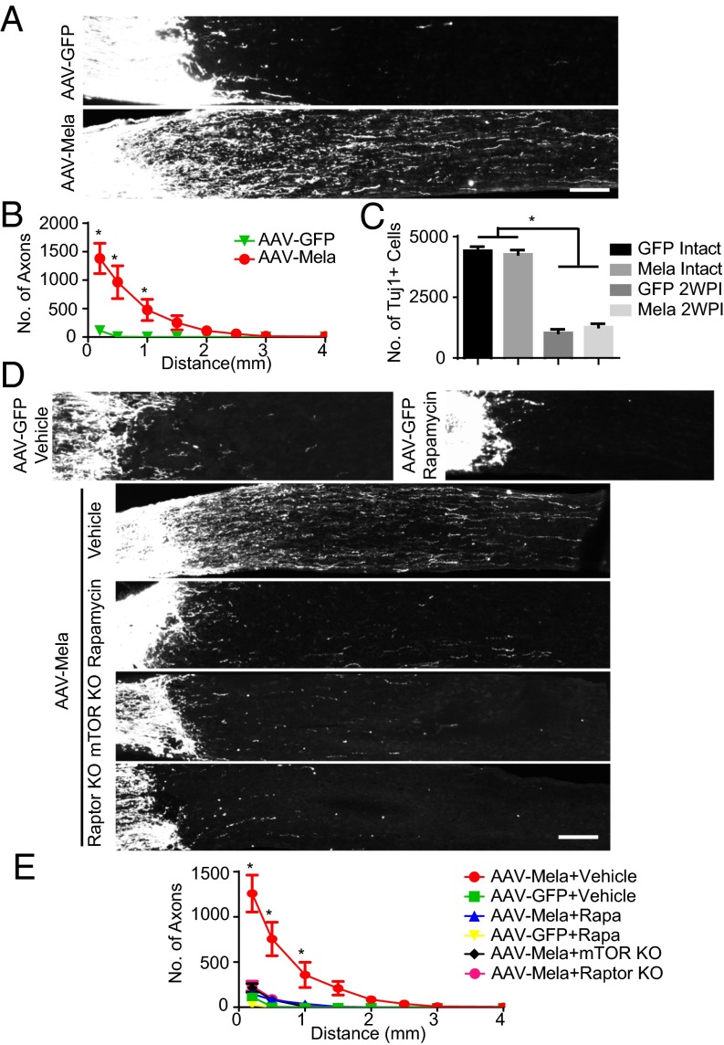
Melanopsin overexpression promotes optic nerve regeneration through an mTORC1-dependent mechanism. (A) Sections of optic nerves containing CTB-labeled axons from WT mice at 2 wk postinjury (2WPI), injected with either AAV-GFP or AAV-Mela. (B) Quantification of regenerating axons at different distances distal to the lesion sites. *P < 0.05, ANOVA followed by Fisher’s LSD, six mice in each group. (C) Quantification of the densities of Tuj1+ RGCs in intact and 2WPI retinas. *P < 0.05, ANOVA followed by Bonferroni’s post hoc test, six mice in each group. (D) Sections of optic nerves from WT, mTOR floxed, and Raptor floxed mice at 2 wk after crush. WT mice were injected with either AAV-GFP or AAV-Mela and administered vehicle or rapamycin (6 mg/kg). Floxed mice were coinjected with AAV-Cre and AAV-Mela. (E) Quantification of regenerating axons from the six groups at different distances distal to the lesion sites. *P < 0.05, ANOVA followed by Tukey’s test, five to six mice in each group. (Scale bars: A and D, 100 μm.)

