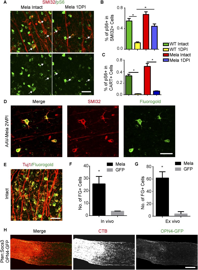
Fig. S6.
Melanopsin promotes axon regeneration of αRGCs. (A) Whole-mount retinas from WT mice injected with AAV-GFP or AAV-Mela with intact optic nerves or 1DPI. Retinas were stained with SMI32 (red) and pS6 (green) antibodies. (Scale bar: 50 μm.) (B) Quantification of percentages of SMI32+ RGCs expressing pS6. (C) Quantification of percentages of cocaine- and amphetamine-regulated transcript-positive RGCs expressing pS6. *P < 0.05, ANOVA followed by Tukey’s test, three to five mice in each group. (D) A whole-mount retina from a mouse with AAV-Mela showing SMI32+ RGCs retrogradely labeled with FG by direct injection into the optic nerve in vivo. SMI32 was positively stained in 95% and 92% of FG+ RGCs for the in vivo and ex vivo labeling, respectively. (E) A whole-mount retina from an intact mouse showing that FG nonselectively labeled Tuj1+ RGCs. (F) For the in vivo labeling, quantification of numbers of FG+ cells per retina in mice injected with AAV-Mela or AAV-GFP. Student’s t test, *P < 0.05, four mice in each group. (G) For the ex vivo labeling, quantification of numbers of FG+ cells per retina in mice injected with AAV-Mela or AAV-GFP. Student’s t test, *P < 0.05, three mice in each group. (H) Images of an injured optic nerve with CTB-labeled axons from Ptenf/f;Socs3f/f;Opn4-GFP mice injected with AAV-Cre and AAV-Mela. GFP+ axons did not regenerate and were not colocalized with CTB+ axons beyond the lesion. (Scale bars: A, D, and E, 50 μm; H, 100 μm.)

