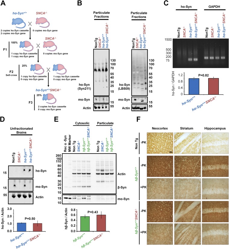
Fig. S6.
Generation and characterization of Tg mice expressing hα-Syn or hβ-Syn in the absence of mα-Syn. (A) Schematic representation of the strategy used to generate Tg mice expressing hα-Syn in the absence of mα-Syn. First, Tg mice expressing hα-Syn (hα-Syn+/+; blue) under the PDGFβ promoter (35) were crossed with SNCA−/− mice (red) (13) to generate heterozygotes (F1). These mice then were backcrossed with SNCA−/− mice before being self-crossed to generate Tg mice expressing two copies of hα-Syn in the absence of any mα-Syn expression (hα-Syn+/+SNCA−/−, F3). (B–D) Brains of age-matched hα-Syn+/+ or hα-Syn+/+ SNCA−/− Tg mice (with non-Tg and SNCA−/− mice as controls) were lysed. Then, protein from particulate fractions (B), total RNA (C), or total proteins in unfractionated brains (D) were extracted. (B) Western blotting of particulate fractions using two different antibodies against hα-Syn [Syn-211 (Left) and LB-509 (Right)] shows increased HMW species in detergent-insoluble fractions of hα-Syn+/+ SNCA−/− Tg brains, compared with hα-Syn+/+ mice and non-Tg and SNCA−/− controls. (C) Semiquantitative RT-PCR using specific primers against hα-Syn or GAPDH (housekeeping gene for normalization) followed by agarose gel electrophoresis demonstrates that no significant differences in hα-Syn mRNA levels were detected between hα-Syn+/+ and hα-Syn+/+ SNCA−/− Tg mice brains. (D) Western blotting of unfractionated brains (homogenized and then centrifuged at 5,000 × g for 5 min) of hα-Syn+/+ or hα-Syn+/+ SNCA−/− mice showed that no significant differences in hα-Syn total protein levels were noted between the two conditions. hα-Syn and mα-Syn were differentially revealed using the Syn211 and D37A6 antibodies, respectively, and actin was used to control for equal protein loading. (E) hβ-Syn+/+ and hβ-Syn+/+SNCA−/− mice exhibit similar β-Syn levels in cytosolic fractions. β-Syn was not detected in particulate counterparts. Loss of mα-Syn expression in SNCA−/− mice is verified using the α-Syn–specific antibody SA-3400 and actin to assure equal protein loading. The Mann–Whitney test was applied in C–E to obtain P values, and three independent experiments were performed. (F) Compared with non-Tg mice, the brains of both hβ-Syn+/+ and hβ-Syn+/+SNCA−/− mice show increased terminal distribution of β-Syn in the neocortex, striatum, and hippocampus, that weakens upon proteinase K (PK) treatment. No hβ-Syn inclusions were noted across conditions. (Scale bar: 100 µm.)

