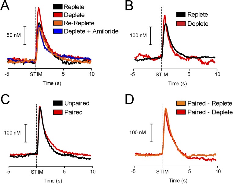
Fig. S5.
In experiments where differences in phasic dopamine release were observed, all recording sites support phasic dopamine release (related to Figs. 1, 2, and 4). Before and after all experimental sessions, we electrically stimulated the VTA (60 Hz, 120 μA, 24 monophasic pulses). Critically, all recording sites supported electrically evoked phasic dopamine release, thereby reducing the possibility that differences in phasic signaling we observed arose from electrodes that were poorly positioned to capture phasic signals. (A) Average phasic dopamine spike during 5 s before to 10 s after electrical stimulation (STIM) of the VTA in rats that received intraoral infusions of NaCl in different states of sodium balance (SEM omitted for clarity because error bars overlap). Experimental data are presented in Fig. 1. Black indicates Replete (n = 4), red indicates Deplete (n = 5), orange indicates Re-Replete (n = 4), and blue indicates Deplete + Amiloride (n = 5). (B) Same as in A but for rats that were trained to associate a CS with the NaCl US while sodium-replete for 7 d and then either depleted of sodium (Deplete, red, n = 5) or given control injections (Replete, black, n = 5). Experimental data are presented in Fig. 2. (C) Same as in A but for rats that received NaCl CS-US training and depletion for 4 d in an unpaired (black, n = 5) or paired (red, n = 5) fashion. (D) Same as in A but for paired rats that were tested while sodium-replete (n = 7) and then again when sodium-deplete (n = 4 of 7). Experimental data from C and D are presented in Fig. 4.

