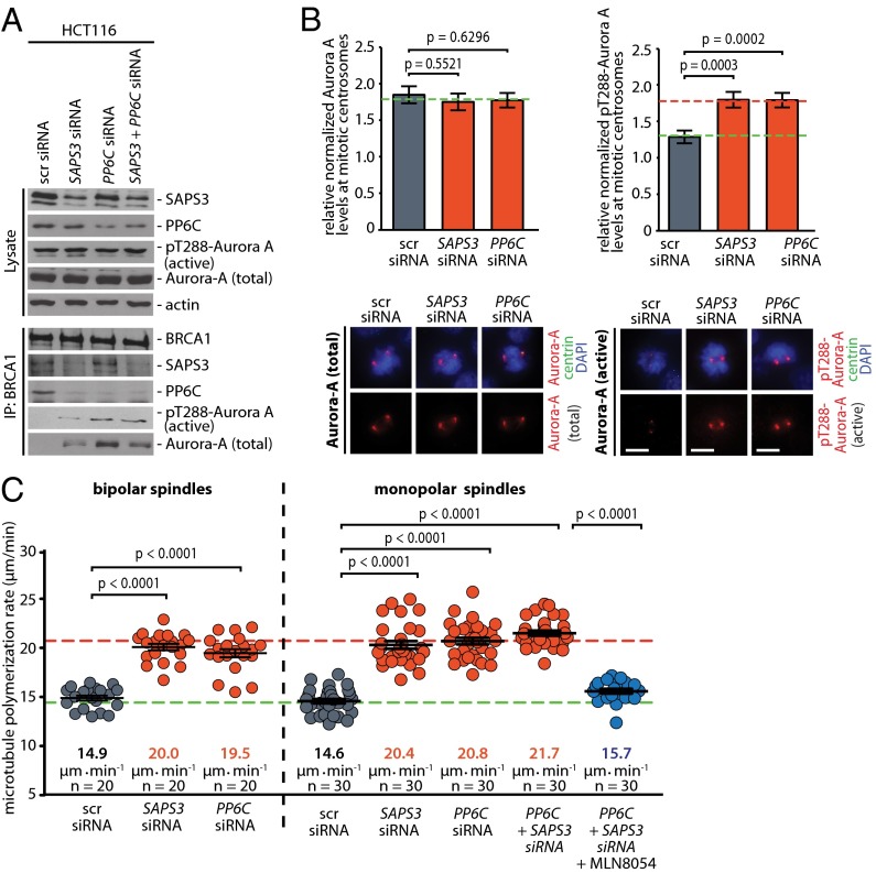
Fig. 2.
Repression of PP6C-SAPS3 is sufficient to increase the activity of centrosomal Aurora-A and accelerates microtubule assembly rates. (A) Repression of PP6C or SAPS3 increases the binding of active Aurora-A to BRCA1. PP6C and/or SAPS3 were repressed in HCT116 cells by siRNAs, and BRCA1 was immunoprecipitated from mitotic cell lysates. The indicated proteins were detected on Western blots. Representative Western blots are shown. (B) Repression of PP6C or SAPS3 increases centrosomal Aurora-A activity. Detection of total or active Aurora-A (pT288) and centrin-2 at mitotic centrosomes in HCT116 cells upon siRNA-mediated repression of PP6C or SAPS3. (Lower) Representative immunofluorescence images of Aurora-A or active pT288-Aurora-A (red), centrin-2 (green), and chromosomes (blue). (Scale bar, 10 µm.) (Upper) The signal intensities were normalized to signals form centrosomal centrin-2 and quantified. Data are shown as mean ± SEM; t test; n = 100 cells. (C) Repression of PP6C or SAPS3 increases mitotic microtubule assembly rates. Microtubule plus-end assembly rates were determined in bipolar metaphase or monopolar spindles after siRNA-mediated repression of PP6C or SAPS3. To inhibit Aurora-A activity partially, mitotic cells were treated with the Aurora-A inhibitor MLN8054 (0.06 µM) for 1 h. Scatter dot plots show average assembly rates (20 microtubules per cell). Green dashed line, normal values; red dashed line, abnormally increased values. Data are shown as mean ± SEM; t test; n = 20–30 cells from three independent experiments.

