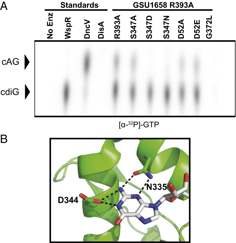
Fig. 3.
Identification of specificity position in Hypr GGDEF active site. (A) Cellulose TLC of radiolabeled products from enzymatic reactions with 1:1 ATP-to-GTP substrates in excess and doped with trace amounts of α-32P-labeled GTP (full TLC and results for α-32P-labeled ATP are shown in SI Appendix, Fig. S10). Residue R393 is located in the putative I-site, S347 is located in the nucleotide binding site, and D52 is the putative phosphorylation site in the Rec domain. (B) Nucleotide binding region of PleD in complex with nonhydrolyzable GTP analog (Protein Data Bank 2V0N, ref. 26). Hydrogen bonding contacts between the guanine base and key protein residues are shown as dotted lines.

