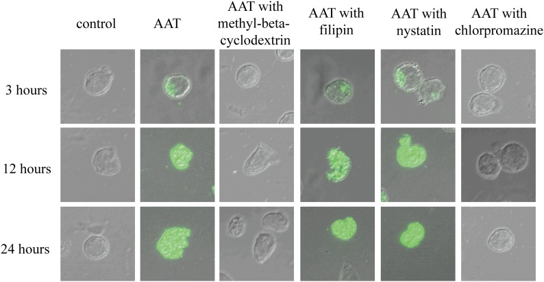
Figure 1. HIV-1-infected CD4+ T cells internalized AAT through a clathrin-dependent endocytosis process.
Activated primary CD4+ T cells were infected with HIV-1IIIB and then cultured with the presence or absence of methyl-β-cyclodextrin, filipin or nystatin, or chlorpromazine (all 20 µg/ml) for 1 h. Next, these T cells were incubated with the presence or absence of 5 mg/ml Alexa Fluor 488-labeled AAT. After 3, 12, or 24 h of incubation, the cells were collected, fixed, and imaged with a confocal microscope (×600).

