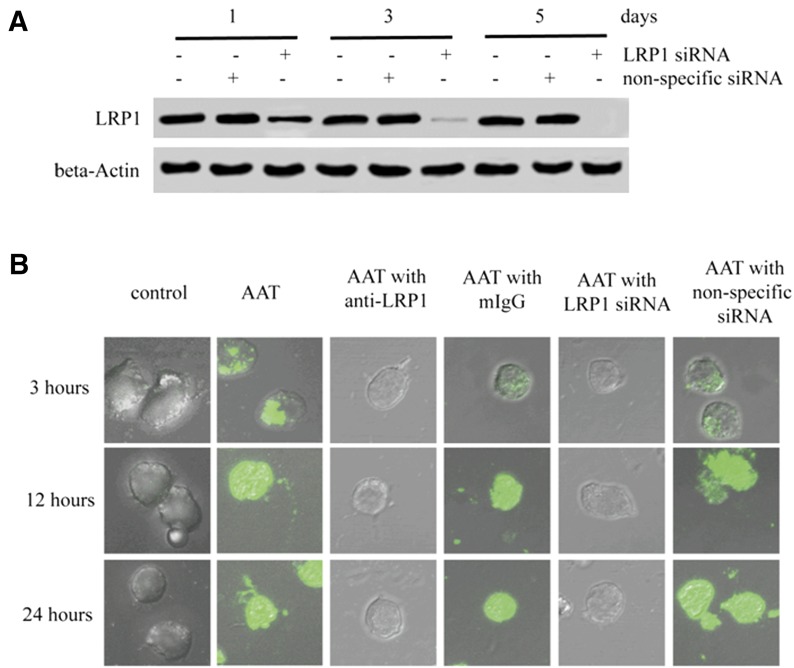
Figure 4. LRP1-mediated AAT internalization in CD4+ T cells.
Activated primary CD4+ T cells were treated with LRP1 siRNA or non-specific siRNA. After 1, 3, or 5 d of incubation, the cells were collected and lysed to detect LRP1 expression by Western blot (A). β-Actin was used a loading control. Moreover, activated CD4+ T cells with or without 3 d of siRNA treatment were infected with HIV-1IIIB and then incubated with the presence or absence of Alexa Fluor 488-labeled AAT, 5 µg/ml LRP1 antibody, or 5 µg/ml mouse IgG for 3, 12, or 24 h. The internalization of AAT was then detected by confocal microscope (B) (×600).

