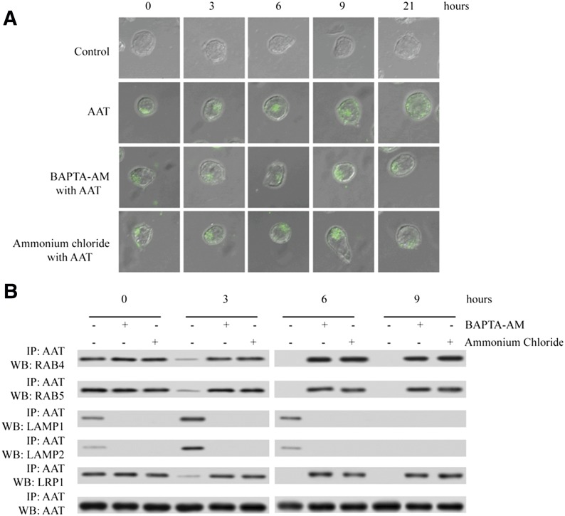
Figure 7. Lysosome inhibitor, NH4Cl, and endosome-lysosome fusion inhibitor, BAPTA-AM, suppressed the transportation of AAT from the endosome to the lysosome.
Activated primary CD4+ T cells were infected with HIV-1IIIB and then cultured with the presence or absence of Alexa Fluor 488-labeled AAT. After 3 h of incubation, unbound AAT was removed, and the cells were treated with the presence or absence of 10 mM NH4Cl or 10 µM BAPTA-AM for another 0, 3, 6, 9, or 21 h. AAT internalization was detected by confocal microscope (×600) (A). The cells after 0, 3, 6, and 9 h of incubation were also treated with DSP to stabilize the protein interaction, and these cells were then lysed to extract the whole cell proteins. AAT antibody was added to the lysate to precipitate the proteins interacting with AAT. LRP1, RAB4, RAB5, LAMP1, AAT, and LAMP2 were detected by Western blot (B).

