Figure 1.
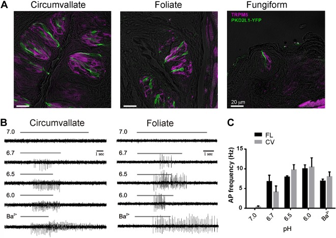
PKD2L1 cells from different parts of the tongue fire action potentials in response to acids. A) Confocal image showing the distribution of PKD2L1 cells identified by expression of YFP under the PKD2L1 promoter in each of the major taste fields on the tongue. YFP (green) and TRPM5 (purple) were detected with protein-specific antibodies, and fluorescent images were overlaid onto a differential interference contrast image of the same field (gray). Note that there is no overlap between the 2 populations. B) Action potentials evoked by solutions of varying pH recorded from dissociated PKD2L1 taste cells from the 2 taste fields indicated using the loose patch recording method. Barium (2 mM) was used as a positive control. Each vertical series is from the same cell except for pH 6.7 in foliate was from a different cell than other pHs. C) Summary data of AP frequency in response to acid stimuli; 4 cells were tested for each condition. No significant differences were found between cells from the CV or FL papillae (2-tailed Student's t test).

