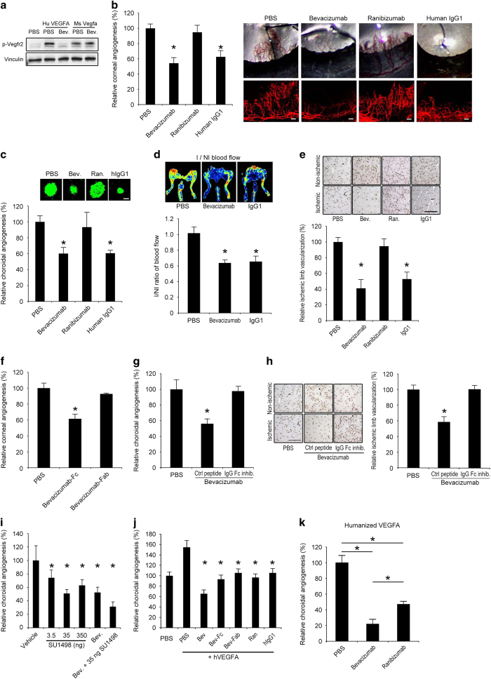
Figure 1.
Bevacizumab inhibited mouse angiogenesis via Fc region. (a) Western blot shows that bevacizumab inhibited Vegfr2 phosphorylation (pVegfr2) in Py4 mouse hemangioma endothelial cells when treated with human VEGFA, but not when treated with mouse Vegfa, after 10 min. Image representative of three experiments. (b) Bevacizumab and human IgG1, but not ranibizumab, decreased corneal angiogenesis in wild-type mice. Area of angiogenesis was measured 10 days after suture injury and normalized to PBS group. n=10–38. Representative photos of wild-type mouse eyes (upper row) and corneal flat mounts (lower row) showing reduced growth of blood vessels (CD31+, red) in eyes treated with bevacizumab or human IgG1, but not in eyes treated with ranibizumab. Scale bars, 100 μm. (c) Bevacizumab and human IgG1, but not ranibizumab, suppressed choroidal angiogenesis in wild-type mice 7 days after laser injury compared with PBS (experiment performed in JA laboratory). Images depict representative choroidal angiogenesis lesions (endothelial cells stained in green) in each treatment group. n=12–20. (d, e) Treatment of ischemic hind limb with bevacizumab or human IgG1 in wild-type mice suppressed muscle revascularization and decreased blood vessel perfusion, as seen in representative laser Doppler perfusion images (top), and measured blood flow in the ischemic limbs (bottom), normalized to the contralateral non-ischemic limbs, 7 days after surgery. n=6. I/NI, ischemic/non-ischemic. Bevacizumab and human IgG1, but not ranibizumab, treatment of ischemic limbs reduced muscle angiogenesis (CD31+, brown) as seen in representative images of muscle CD31 immunolocalization (e), and quantification of muscle CD31 immunolocalization (bottom), normalized to the contralateral non-ischemic limbs. (f) The Fc fragments, not the Fab fragment, of bevacizumab suppressed corneal angiogenesis in wild-type mice. Area of angiogenesis was measured 10 days after suture injury and normalized to PBS group. n=10–38. (g) Co-administration of a peptide that prevents IgG-Fc binding to FcγR, but not a control peptide, blocked inhibition of choroidal angiogenesis by bevacizumab in wild-type mice. (h) Co-administration of an IgG-Fc inhibitory peptide, but not a control peptide, blocked inhibition of muscle angiogenesis (CD31+, brown) by bevacizumab, as seen in representative images of muscle CD31 immunolocalization (left), and quantification of muscle CD31 immunolocalization (right), normalized to the contralateral non-ischemic limbs. Scale bar, 100 μm. n=6. (i) Bevacizumab suppressed choroidal angiogenesis in wild-type mice to the same extent as SU1498, a small molecule tyrosine kinase inhibitor of Vegfr2. Combined administration of bevacizumab and SU1498 suppressed choroidal angiogenesis to a greater extent than either of the agents alone. n=6. (j) Choroidal angiogenesis, augmented by administration of human VEGFA, was suppressed to similar extents by ranibizumab, bevacizumab-Fab, bevacizumab-Fc and human IgG1; and, to a greater extent, by bevacizumab. n=6–8. (k) Bevacizumab suppressed choroidal angiogenesis to a greater extent than ranibizumab in the humanized VEGFA mouse, a transgenic model that expresses a VEGFA protein that can be neutralized by both bevacizumab and ranibizumab. n=6. Results are means±s.e.m. *P<0.05 compared with PBS (b–h, k) or with vehicle (i) or with PBS+human VEGFA (j).

