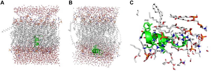
Figure 4.
A) Starting structure of the orientation of Cm-p5 (green helix) embedded in the hydrophobic area of the phosphatidylserine bilayer. On top and bottom, surrounding water layers are displayed. B) Interaction of the Cm-p5 (green helix) with the surface of the membrane bilayer model. C) Details of the interaction of the Cm-p5 (green helix) with head groups of phosphatidylserine.

