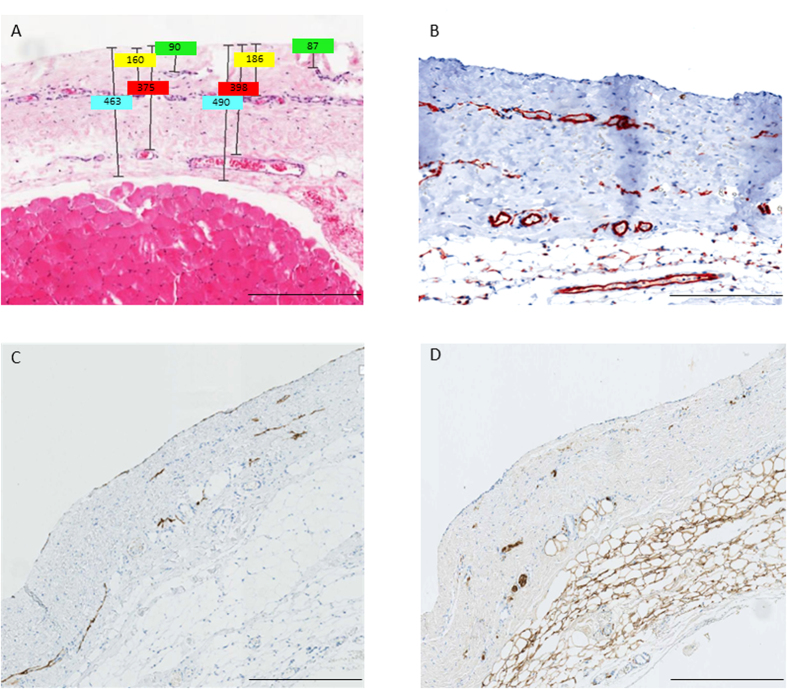
Figure 2.
Hematoxylin-eosin (A) and CD31 (B) stainings of parietal peritoneum demonstrating the three layer structure (A,B) and co-localization of podoplanin positive lymphatic capillaries (C) and S100A4 positive nerve fibers (D). Scale bars: 400 μm. Illustrative examples of measurements of submesothelial thickness, and of the distance of the three vessel layers to the mesothelial surface are given in Fig. 2A.

