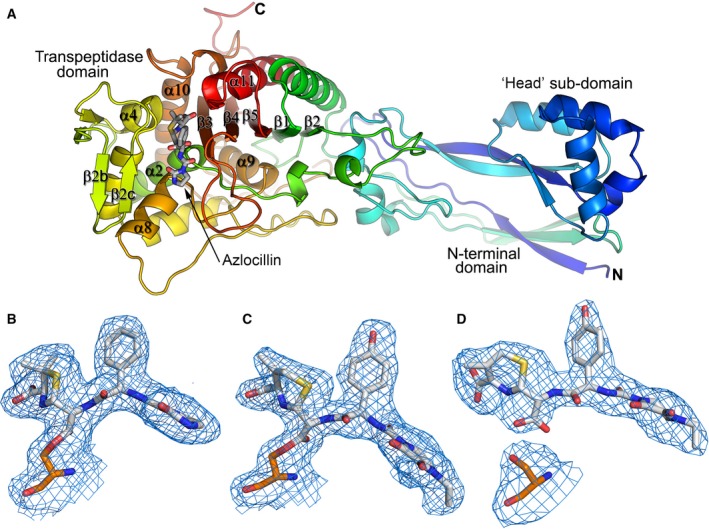
Figure 2.

Overall structure of PBP3 and electron density maps. (A) Rainbow coloured cartoon representation of azlocillin PBP3 AEC, showing the overall structure of PBP3 and penicillin‐binding site. (B–D) F o ‐F c omit electron density maps contoured at 3σ showing covalent binding of (B) azlocillin and (C) cefoperazone to give acyl–enzyme complexes, and noncovalent binding of (D) anhydrodesacetyl cephalosporoic, the product of deacylated cefoperazone, at the active site of PBP3. The antibiotics are shown with grey bonds and S294 is shown with orange bonds.
