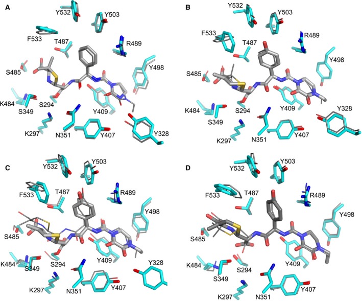
Figure 4.

Comparison of covalent and noncovalent PBP3 complexes (A) Superposition of PBP3–azlocillin and PBP3–piperacillin (PDB id 4KQO) covalent complexes, (B) PBP3–cefoperazone and PBP3–piperacillin covalent complexes. (C) PBP3–cefoperazone and PBP3–anhydrodesacetyl cephalosporoic (ACA) complexes and (D) PBP3–ACA and PBP3–PA (PDB id 4KQR) complexes. In (A–C), the protein side‐chains in the azlocillin and cefoperazone acyl complexes are shown as thick cyan sticks and β‐lactams as thick grey sticks, the inhibitor and protein side‐chains in the piperacillin acyl complex and the PBP3‐ACA complex are shown as thinner sticks. The side‐chains and the inhibitor of PBP3‐ACA complex are shown as thick sticks in (D).
