Fig. 5.
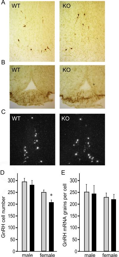
GnRH is not affected with cFOS deficiency. A, Immunohistochemistry of the preoptic area with GnRH antibody was used to identify GnRH neurons in the hypothalami of 6-week-old wild-type (WT) and null (KO) mice. B, Median eminence was stained for GnRH. C, In situ hybridization was performed to analyze GnRH neurons and quantification of neuron number presented as the group mean ± SEM of 5 animals per group in D, while quantification of the grains per cells to analyze mRNA expression is presented in E. Light bars, wild type; black bars, cFOS nulls in D, E. Results of quantifications were presented as the mean ± SEM and asterisks (*) indicate significant difference (p < 0.05) in null females in GnRH neuron number, as determined by two-factor ANOVA and Tukey–Kramer HSD post hoc test.

