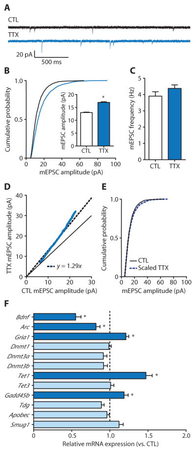
Fig. 1. Homeostatic upscaling of excitatory strength is associated with alterations in mRNAs for gene products associated with the regulation of DNA methylation.
(A) Sample mEPSC records from cortical pyramidal neurons after 24 hours of exposure with control (CTL) or TTX (blue). (B and C) Cumulative probability distributions and mean mEPSC amplitudes (B) and mean mEPSC frequencies (C) from cortical pyramidal neurons treated with TTX. Bar graphs are means ± SEM from cells pooled from at least six experiments for each condition (CTL, n = 21 cells; TTX, n = 15 cells). (B) P < 0.001, Kolmogorov-Smirnov (K-S) test. Inset: *P < 0.001, Mann-Whitney (M-W) test. (D) Rank order plot of 1000 randomly selected mEPSC amplitudes from CTL and TTX (blue values). Fitting the data with y = ax (dashed black line) yielded a slope of 1.29, which was used to scale down amplitudes from TTX-treated cells in (B). The solid black line represents unity. (E) Cumulative probability distributions of scaled-down TTX-induced amplitudes compared to those from CTL cells; P = 0.1024, K-S test. (F) Polymerase chain reaction (PCR) detection of transcript expression relative to CTL after 24-hour TTX treatment. Dashed vertical line represents the average, normalized gene expression values of CTL cells. Data are means ± SEM from three experiments. *P < 0.05, Student’s unpaired t test.

