. 2015 Oct 21;5(1):188–196. doi: 10.1039/c5tx00164a

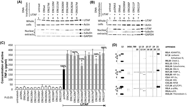
Fig. 4. WB/ELISA analysis of serine mutation of ERK2. To examine the effect of serine on LITAF regulation, western blot was performed (A & B). RAW 264.7 cells were untreated (lanes 1 & 11) as the control or co-transfected with LITAF and ERK2 cloned DNAs (lanes 2–9, 12–17). Cell extractions from whole cells or nuclei of either control or test groups were separately purified and subjected to Western blot analysis with antibody against LITAF, compared to Actin, tubulin, or GAPDH as the control. Triplicate assays above were conducted. Mean SEM. To further identify which serine is involved in TNF-α secretion via LITAF, ELISA analysis was performed (C). Pre-cultured Raw264.7 cells were untreated as the control (lane 1) or transfected with 0.5 μg ml–1 of ERKwt (lanes 3 & 15), ERKw27M (lanes 4 & 16), ERK120M (lanes 5 & 17), ERK151M (lanes 6 & 18), ERK206M (lanes 7 & 19), ERK244M (lanes 8 & 20), ERK282M (lanes 9 & 21), ERK357M (lanes 10 & 22), ERK206P (lanes 11 & 23), ERK211P (lanes 12 & 24), ERK221P (lanes 13 & 25), or pcDNA3 (lanes 2 & 14) as control. 0.5 μg ml–1 LITAF was simultaneously transfected (lanes 14–25) for 16 h. The supernatants were collected from each test group and used for assessment of TNF-α production with triplicate ELISAs. To detect whether other factors or kinases are affected in the absence of S206 of ERK, a protein array was performed (D). The matured THP-1 cells were co-transfected with 0.5 μg ml–1 LITAF DNA plus 0.5 μg ml–1 of pcDNA3 (filter I) as the control or ERK2WT (filter II) or ERK206P (filter III) for 48 h. The protein from each treated cell was purified and used for protein array assay by a Human Cell Stress Array Kit (R & D Systems ARY018) following manufacturer's instruction.

Fig. 4