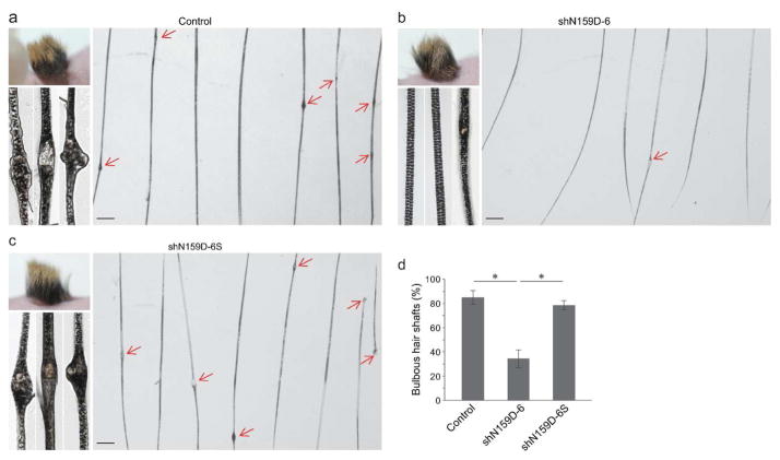
Figure 2. Phenotypes of hair regenerated with shRNA-modified homozygous mutant Krt75 keratinocyte progenitor cells.
(a–c) Representative gross appearance, low and high power images of hair regenerated with non-infected cells. Control (a), shN159D-6 lentiviral vector infected cells (b), and scrambled (shN159D-6S) lentiviral vector infected cells (c) at one month of grafting. Arrows point to bulbous lesions (blebs) along the hair shaft. (d) Quantification of hair shafts containing blebs. Asterisk, P < 0.05. Scale bar: 250 μm.

