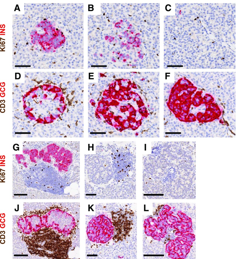
Figure 2.
Heterogeneity of islet β-cells and the numbers of CD3+ cells in insulitic islets in young donors at diabetes onset and after 5 years of disease duration. Representative islets were imaged for a 13-year-old at diabetes onset (nPOD 6228; A–F) and for another 13-year-old with T1D for 5 years (6243; G–L). Serial paraffin sections were stained using dual-IHC (Ki67 plus insulin [INS], CD3 plus glucagon [GCG]), as described in research design and methods. Decreasing proportions of β-cells are shown in different islets from both donors (A–C and G–I) as well as heterogeneity in numbers of CD3+ cells per islet (D–F and J–L). Heterogeneity in CD3+ counts between two insulin− islets of similar sizes is also shown for one donor (K and L). Scale bars: A–F, H, and K, 50 μm; G, I, J, and L, 100 μm.

