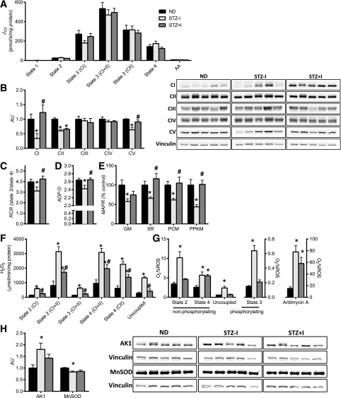
Figure 1.
Insulin deprivation adversely affects skeletal muscle mitochondrial physiology. A: Oxygen consumption (JO2) was not different across groups. Expression of mitochondrial respiratory CI and V proteins (B) and mitochondrial coupling efficiency, measured by respiratory control ratios (RCR) (C) and ADP:O ratio (D), was reduced by insulin deprivation and corrected by insulin treatment. E: Mitochondrial ATP production rate (MAPR) was significantly lower with insulin deprivation and restored with insulin therapy. F: Mitochondrial H2O2 emissions were elevated with insulin deprivation and partially reduced by insulin replacement across all respiratory states. G: O2 consumption as a percent of ROS was higher in STZ–I. H: Increase in mitochondrial ROS production was associated with downregulation of mitochondrial MnSOD. Increased expression of AMP-sensitive adenylate kinase 1 (AK1) confirms the low-energy state of skeletal muscle in insulin-deprived animals. Substrate combinations description: GM, glutamate and malate; SR, succinate and rotenone; PCM, palmitoylcarnitine and malate; PPKM, palmitoylcarnitine, pyruvate, α-ketoglutarate, and malate. Data are means ± SEM (n = 13 per group for mitochondrial functional measurements, n = 6 for Western blot measurements). AA, amino acids; AU, arbitrary unit. *P < 0.05 vs. ND; #P < 0.05 vs. STZ–I group.

