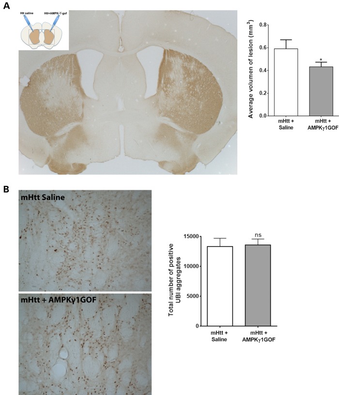
Figure 6.
In vivo activation of AMPK rescues striatal cell degeneration in a mouse model of HD. (A) Diagram showing the treatment of both sides of the brains of mice with lentiviruses expressing the N-terminal fragment of mHtt [Htt171-82Q (Htt in the diagram)], or the AMPKγ1GOF. Below is shown a representative picture of DARPP32 staining of the sections of the brains of these mice, which highlights the area of the lesion caused by mutant Htt. The right panel shows the analysis of the volume of the lesion caused by mutant Htt, with overexpression of AMPKγ1GOF reducing cell death. Values are mean ± SEM. *P < 0.05 (P = 0.03, N = 13). (B) Representative pictures showing UBI staining and pointing out to inclusion bodies. The right panel shows the quantification of the number of UBI-positive aggregates. Values are mean ± SEM. The overexpression of AMPKγ1GOF does not change the number of inclusion bodies (P = 0.87, N = 13). Statistics use the Wilcoxon test for paired samples. ns: not significant.

