Figure 2.
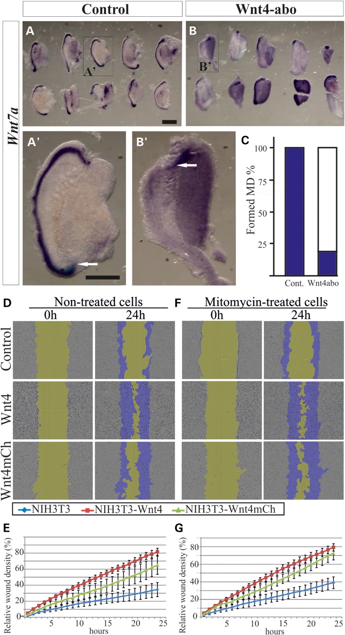
The Wnt4 function is required for the extension of the MD and scratch-wound recovery in vitro. Urogenital ridges were dissected at E11.5 and grown for 48 h in the presence of goat IgG immunoglobulins as controls (A). The MD was detected with the Wnt7a marker (A, and enlarged in A′, arrow). The MD failed to develop in ∼79% of the cases upon supplementation with anti-Wnt4 antibody (B, and enlarged in B′, arrow) (C). (D) Micrographs from the in vitro wounding assay with control NIH3T3, NIH3T3Wnt4+ and NIH3T3Wnt4mCh/mCh+ cells reveal a positive influence of Wnt4 on wound closing, as quantified in (E). (F) Treatment of the cells with mitomycin did not alter the behaviour of these cells significantly, as quantified in (G). Scale bars (A, B and A′–B″) 500 µm.

