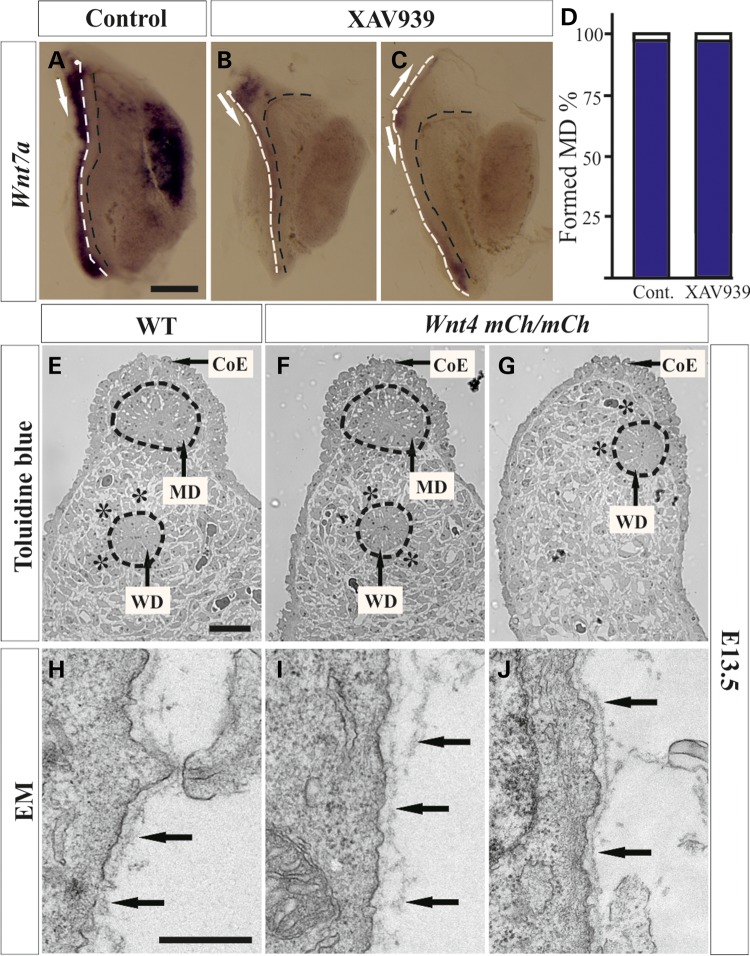
Figure 3.
Changes in MD growth caused by the tankyrase inhibitor XAV939 and the hypomorphic Wnt4mCh/mCh allele. The MD develops normally in the presence of DMSO, serving as a control (A, white line, arrow). The Wnt7a MD marker is weak in the presence of XAV939, so that ∼41% of the MDs have branched on the apical side (B, arrows) and some have extended not only posteriorly, but also anteriorly (C, arrows). XAV939 does not block MD growth, but it severely affects the properties of the resulting MD (D). (E) Toluidine blue staining detects the wild-type urogenital ridge at E13.5. The MD was formed in 45% of the Wnt4mCh/mCh mice (F) and failed to form in 55%. The mesenchymal cells were not organized in a concentric manner around the WD at E13.5 in the Wnt4mCh/mCh mice, in contrast to the controls (stars in E and G). The failure of MD formation influences the localization of the WD and CoE (arrows in E and G). The transmission electron micrographs depict a well-condensed BM around the control MD (H, arrows) but a loose, malformed one around its Wnt4mCh/mCh counterpart (I, arrows). The BM surrounding the WD remains intact (J, arrows). MD, white dashed line, and WD, black dashed line in (A–C). CoE, coelomic epithelium. Scale bars (A–G) 500 µm and (H–J) 250 nm.

