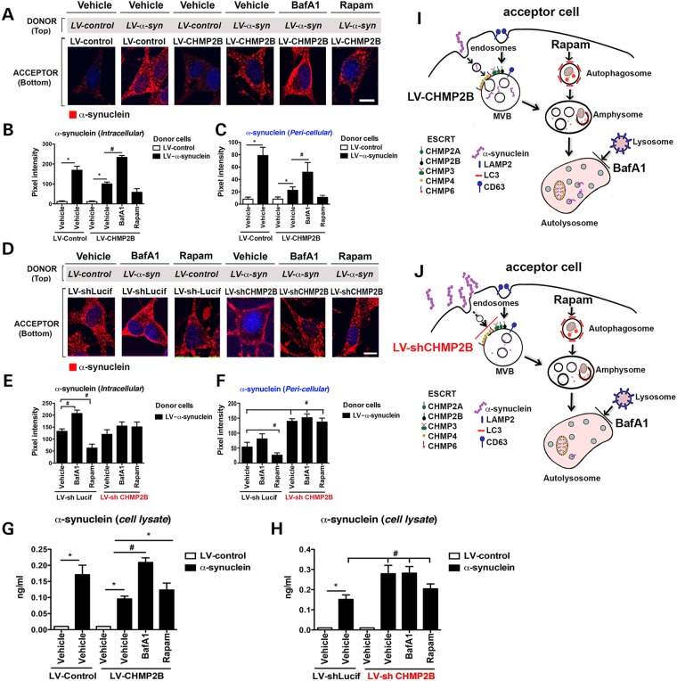
Figure 3.
Endocytosed α-syn is transported to the autophagosome for degradation in an ESCRT-III-dependent manner. (A) Immunohistochemistry of acceptor cells infected with LV-control or LV-CHMP2B co-cultured with donor cells infected with LV-control or LV-α-syn. Cultures were then treated with BafA1, Rapamycin (Rapam) or Vehicle. Coverslips were stained for α-syn (red) and nuclei (DAPI, blue). (B and C) Coverslips were analyzed to determine levels of α-syn immunoreactivity expressed as pixel intensity. (D) Immunohistochemistry of acceptor cells infected with LV-shCHMP2B or LV-shLucif co-cultured with donor cells infected with LV-α-syn. Cultures were then treated with Baf, Rapam or Vehicle. Coverslips were stained for α-syn (red) and nuclei (DAPI, blue). (E and F) Coverslips were analyzed to determine levels of α-syn immunoreactivity expressed as pixel intensity. (G and H) Lysates from acceptor cells were assayed by electrochemical α-syn assay. (I) Model of the effects of CHMP2B overexpression on α-syn autophagy degradation. (J) Model of the effects of CHMP2B downregulation on α-syn autophagy degradation. *Indicates statistical significance P < 0.05 compared with vehicle treatment with LV-control-infected donor cells. #Statistical significance P < 0.05 compared with vehicle treatment with LV-α-syn-infected donor cells. One-way ANOVA with post hoc Tukey–Krammer (n = 3 experiments per group).

