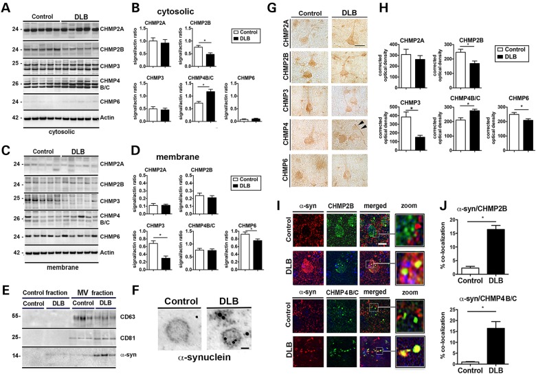
Figure 5.
Dysregulation of ESCRT-III proteins in DLB. Western blot analysis of (A) cytosolic and (C) membrane fractions of brain homogenates showing levels of ESCRT-III complex proteins CHMP2A, CHMP2B, CHMP3, CHMP4B/C and CHMP6 on postmortem human brain samples from control subjects and DLB patients. Densitometric analysis of (B) cytosolic and (D) membrane fraction western blots of CHMP2A, CHMP2B, CHMP3, CHMP4B/C and CHMP6 immunoreactivity analyzed as ratio to β-actin signal. (E) Microvesicles (MV) isolated from whole brain homogenates of normal or DLB patient brains were analyzed by immunoblot for the multivesicular body marker CD63 and CD81 as well as the presence of α-syn. (F) Electron microscopy of isolated MV stained with gold particle labeled anti-α-syn. Scale bar = 50 nm (n = 4 control and n = 4 DLB cases). (G) Immunohistochemical detection of CHMP2A, CHMP2B, CHMP3 and CHMP4B/C in control and DLB brains. Scale bar = 20 µm. Arrowheads indicate Lewy neurite. (H) Optical density analysis of immunohistochemical immunoreactivity. (I) Brain sections from the control subject and DLB patients were double labeled with antibodies against α-syn (red) and CHMP2B or CHMP4B/C (green) and imaged with the laser scanning confocal microscope. (J) Analysis of % of cells showing co-localization between α-syn and CHMP2B or CHMP4B/C. *Indicates statistical significance P < 0.05 compared with control subjects. One-way ANOVA with post hoc Tukey–Krammer (n = 8 control cases and n = 12 DLB cases).

