
Fig. 1.
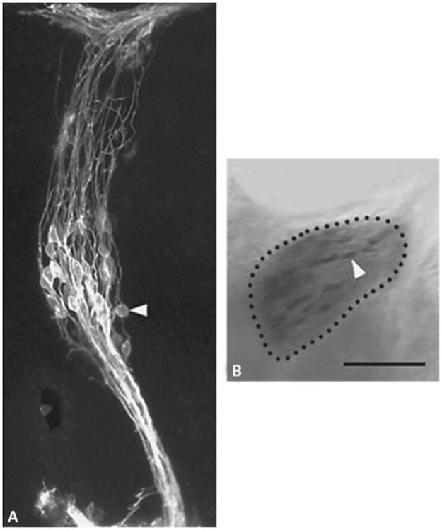
Geniculate neurons labeled by DiI and following photoconversion of DAB. A A projection of optical sections obtained from a Zeiss 410 confocal microscope showing individual neurons labeled 24 h after application of DiI to the PA/CT of a fixed E11 mouse geniculate ganglion. B A different ganglion from that shown in A following photoconversion imaged on a conventional Zeiss Axiovert 100 microscope. Bar = 100 μm.