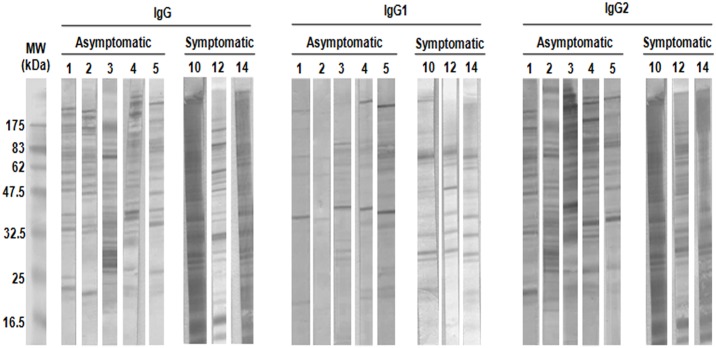
Fig 1. IgG reaction pattern associated with CVL status.
Total Leishmania infantum promastigote cell extracts were separated with 12% SDS-PAGE and electrotransferred to nitrocellulose membranes. The membranes were probed with serum obtained from individual asymptomatic dogs (1:500) or symptomatic dogs (1:1,000) and secondary anti-dog (A) IgG, (B) IgG1 or (C) IgG2 (1:10,000). Representative western blots from each group of dogs are depicted (asymptomatic: n = 5; symptomatic: n = 3). Numbers represent the code numbers of the dogs enrolled in the study. Molecular weight markers are shown on the left-hand side.

