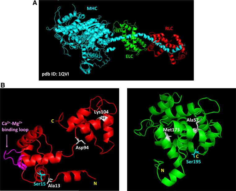
Fig. 1.
a Structure of myosin head (S1) and the lever arm domain (pdb ID: 1QVI); b ITASSER predicted computational structures of human ventricular RLC (left) and ELC (right). Depicted: HCM/DCM mutations in RLC/ELC discussed in this report; phosphorylatable serines, Ser15-RLC and Ser195-ELC; Ca2+-Mg2+ binding site on RLC

