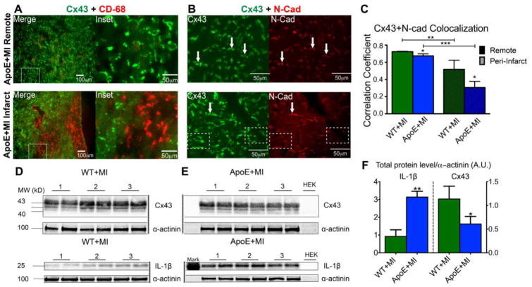
Figure 2.
Inflammation and Cx43. A) Immunofluorescence images of Cx43 (green) and CD-68 (red, macrophage marker) show abundant macrophage infiltration in the infarct region of an ApoE+MI heart (bottom). Myocytes adjacent to macrophages show internalization of Cx43 (Inset). B) Top: Cx43 (green) normally co-localizes with N-cadherin (N-Cad, red), a marker of the intercalated disc. Arrows indicate examples of obvious co-localization of Cx43 and N-Cad. Bottom: In the infarct region of an ApoE+MI heart, Cx43 expression is observed without corresponding N-Cad staining, indicating Cx43 internalization or lateralization to areas outside of the intercalated disc. Arrows indicate obvious examples of co-localization. Dashed boxes indicate examples of internalization (Cx43 without corresponding N-Cad). C) Quantification of Cx43 and N-Cad co-localization, demonstrating increased Cx43 internalization/lateralization in the infarct compared to remote regions, and in ApoE+MI compared to WT+MI hearts. D–F) ApoE+MI hearts have a ~3-fold increase in IL-1β expression and a corresponding ~2-fold decrease in Cx43 expression. Reprinted with permission from [47].

