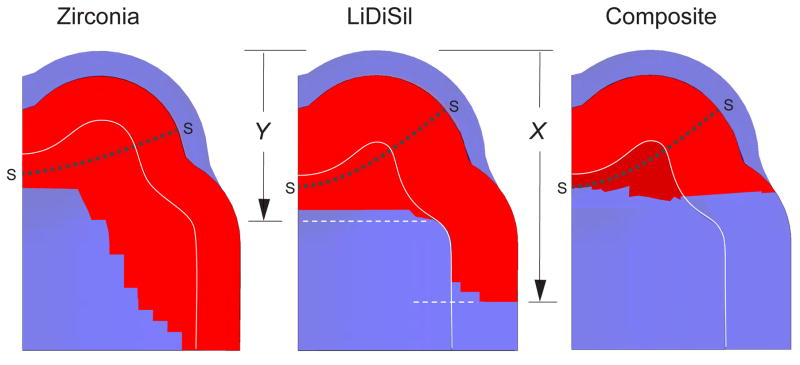
Fig. 3.
XFEM predictions of molar crown splitting for model system in Fig. 1, showing half-sections at point of failure. Calculations made using parameters in Table 1, for crown thicknesses d = 1.0 mm (zirconia) and 2.0 mm (lithium disilicate and dental nanocomposite), base radius R = 5 mm and occlusal-to-margin height H = 7.0 mm, loaded axially with indenting sphere of radius r = 3.2 mm. The sections indicate cracking from starter cracks SS in both the external crown and internal support material, cracks marked in red. Rear cusp lies behind plane of diagram.

