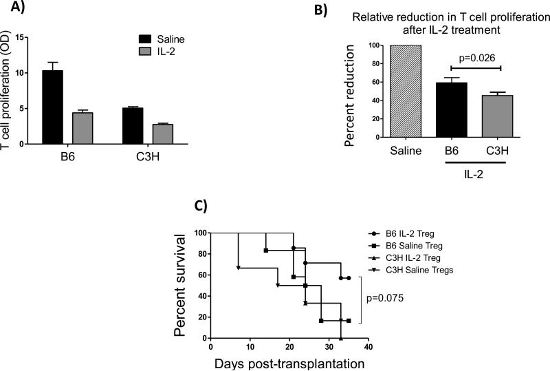
Figure 5. IL-2 expanded Tregs show allo-antigen specificity in vitro and in vivo.
(A, B) Treg suppression assay showing proliferation of naïve CD4+CD25−T cells in the presence of CD4+CD25+Tregs isolated from IL-2 treated vs. saline injected mice in the presence of B6 (donor type) or C3H (third party) APCs (N=5 mice/group), data represents results of two independent experiments. (C) Adoptive transfer of Tregs from IL-2 or saline injected BALB/c recipients of B6 donors to a new set of hosts (N=7/group). Tregs were isolated using flow sorting at 3 weeks post-transplantation; 80,000 Tregs per recipient were i.v. transferred to two different groups of hosts (BALB/c mice receiving transplants from either B6 or C3H donors) 18 hour post-surgery. Graft survival was monitored up to 5 weeks post-transplantation, which revealed an increase survival when hosts were injected with Tregs from IL-2 treated group with prior encounter to the same donor antigen (n = 6-7/group, Kaplan-Meier survival analysis).

