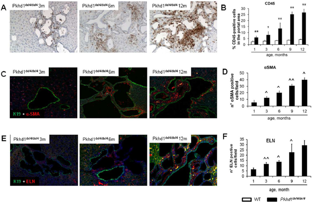
Figure 2. The progressive peribiliary accumulation of inflammatory cells in the portal tract, co-expressing F4/80 (macrophage marker) contrasts with the initial scarce contribution of myofibroblasts.
A. A progressive portal accumulation of CD45+ inflammatory cells adjacent to biliary cysts was observed (immunoperoxidase for CD45, M=200×). B. Amount of CD45+ cells was quantified by morphometric analysis (n=3 for each age). C. In Pkhd1del4/del4 mice, α-SMA+ myofibroblasts scattered within the portal space (dual immunofluorescence for α-SMA - in red, and K19 - in green, M=200×). D. The number increased slowly through maturation with a linear pattern (n=3 for each age). E. Similarly to α-SMA, elastin (ELN)+ portal fibroblasts accumulated into the portal space in a timedependent fashion (dual immunofluorescence for ELN - in red, and K19 - in green, M=200×). F. As quantified by morphometry, the number of ELN+ cells augmented through maturation, with the most relevant increase after 6 months (n=4 for each age). *p<0.05 vs WT (same age), **p<0.01 vs WT (same age); ^p<0.05 vs previous maturation age, ^^p<0.01 vs previous maturation age.

